Long-Term In Vivo Monitoring of Injury-Induced Brain Regeneration of Adult Zebrafish Using Spectral Domain Optical Coherence Tomography

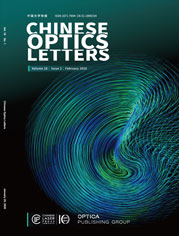
A brain injury model was induced by needle insertion on the head of an adult zebrafish. Long-term in vivo noninvasive monitoring and 3D imaging of the brain regeneration process of the adult zebrafish were successfully achieved using spectral-domain optical coherence tomography (SD-OCT).
The zebrafish, as a model of vertebrate biology, is widely recognized as an excellent system for exploring brain regeneration. To date, few neuroimaging modalities have been able to effectively and noninvasively visualize the structural and functional information of the adult zebrafish brain after injury in real time with high spatial resolution and good penetration depth.
For the first time, to the best of their knowledge, Professor Zhen Yuan's group at the Faculty of Health Science, University of Macau employed a spectral-domain optical coherence tomography (SD-OCT) system for long-term in vivo monitoring of tissue regeneration in three dimensions using a model of an adult zebrafish with brain injury. This pilot work, together with the pronounced regenerative capacity of the adult zebrafish, provides us with an insight into the process of brain regeneration. This report was published in Chinese Optics Letters Volume 14, No.8, 2016 (J. Zhang et al., Long-term in vivo monitoring of injury induced brain regeneration of the adult zebrafish by using spectral domain optical coherence tomography).
Compared with the current imaging techniques, SD-OCT is able to effectively and noninvasively monitor brain regeneration at different stages in a mechanically damaged brain of adult zebrafish in real time. Based on the generated images of the brain, SD-OCT enables the characterization and evaluation for describing the process of brain regeneration with high, three-dimensional spatial resolution (12 μm axial and 13 μm lateral) at video rate. This unprecedented technique using SD-OCT will prove to be a useful tool for studying the zebrafish brain, particularly in biological procedures in vivo and dynamic processes.
"The present work will definitely play an essential role in the investigation of the brain structure and function using an adult zebrafish as the model," said Prof. Wei Ge, the associate dean of Faculty of Health Sciences, University of Macau.
Further work will be focused on performing angiography and molecular imaging on zebrafish head using SD-OCT, and applying these techniques to different biomedical problems.
频域光学相干成像实现成年斑马鱼损伤大脑再生过程的长期活体监控

图片说明:通过将注射针头插入成年斑马鱼头部造成的大脑损伤模型。基于频域光学相干成像(SD-OCT)成功地实现了活体长期非入侵监控和三维成像成年斑马鱼大脑再生过程。
作为脊椎动物,斑马鱼已成为一种非常适合用于大脑再生研究的模型动物,相关研究的关键性需求是精确可视化斑马鱼大脑的结构。到目前为止,尚未有方法能在保证较高分辨率和足够成像深度的前提下,有效地非入侵地对损伤后的成年斑马鱼大脑实时成像。
澳门大学健康科学学院袁振教授课题组首次(据该课题组所知)基于一套长视距频域光学相干成像(SD-OCT)系统,开展系列斑马鱼大脑成像的研究。他们实现了SD-OCT活体三维成像成年斑马鱼大脑损伤以及长期非入侵监控后期的大脑再生过程。相关研究成果发表在 Chinese Optics Letters 2016年第8期上(J. Zhang et al., Long-term in vivo monitoring of injury induced brain regeneration of the adult zebrafish by using spectral domain optical coherence tomography)。
相对于现有的斑马鱼大脑成像技术,SD-OCT能高效地和非入侵地实时监控大脑机械损伤后恢复的各个阶段。其成像质量和成像深度都非常好,足以在微米分辨率(轴向分辨率12 μm、 横向分辨率 13 μm)的条件下以视频速率提供完整成年斑马鱼大脑的三维信息,支持对大脑恢复状态进行全面地评估。这种无与伦比的能力使得SD-OCT有望成为非常有用的斑马鱼大脑研究工具,尤其适合用于一些生物学程序以及动态过程的活体研究。
澳门大学健康科学学院副院长葛伟教授评价说:“这项工作对于以斑马鱼为模型进行大脑结构和功能分析的研究者有着极为重要的价值”。
后续的工作主要集中在两个方面:基于SD-OCT开展斑马鱼大脑血管造影和分子成像研究,以及将SD-OCT具体应用到不同的生物医学难题。

